
")
")
Age Related Macular Degeneration
What is it?
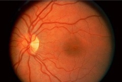
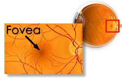
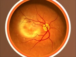
The macula is located in the center of the fundus of the eye, near the optic nerve and is responsible for our central vision (fig.1,2).
 |
 |
After reaching a certain age -according to statistics, over 60- some age-related changes may occur due to the degeneration process. This very serious disease threatens our central vision. We distinguish two types: dry macular degeneration and wet macular degeneration.
A. Dry AMD
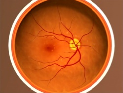
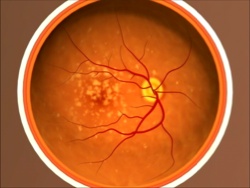
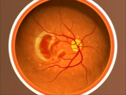
It is the most common lesion. The damage develops gradually and, unfortunately, the patient discovers it at an advanced stage when the loss of vision is already irreversible. Gradually, the macula atrophies or it is destroyed by metabolic deposits, known as Drusens. (fig. 4)
 |
 |
B. Wet AMD
It is a more advanced and damaging form of the disease. The damage develops rapidly because of bleeding or hemorrhage of pathological - fragile - new blood vessels that grow beneath the retina. Patients usually go to the doctor after a sudden loss of vision.
 |
 |
Prevention - Treatment
At the age of 60 years, every patient must undergo examination by an ophthalmologist, who will inform him about his fundus condition. Examination of the fundus is carried out using mydriatic drops (fundoscopy). In case of sudden loss of vision or occurrence of any unusual symptoms, the patient should immediately consult a doctor, regardless of age. The purpose of any treatment is to stabilize patient`s condition.
This severe form of age-related macular degeneration can be treated with:
- Injections (satisfactory, but not permanent outcome),
- Less frequently with laser therapy along with or without injections
In dry form of the disease, we also try to stabilize patient's condition with some medications in the form of antioxidant vitamins and food additives. Thus, preventive examination is very important for accurate and early diagnosis, which contributes to stabilization of the age-related macular degeneration.
Digital content: Credit to National Eye Institute, National Institutes of Health.